Home
Uncategories
Pelvic Anatomy Female / Female Pelvic Anatomy Medical Illustration Medivisuals / The mons pubis, or public mound, is the fleshy area on the pelvic.
Pelvic Anatomy Female / Female Pelvic Anatomy Medical Illustration Medivisuals / The mons pubis, or public mound, is the fleshy area on the pelvic.
Pelvic Anatomy Female / Female Pelvic Anatomy Medical Illustration Medivisuals / The mons pubis, or public mound, is the fleshy area on the pelvic.. Mar 5, 2020 view item. The following sections discuss these in more detail. The female pelvis was designed to make the subject of the pelvis understandable to all women. (2017, elsevier) should be consulted. 1 chapter 1 applied clinical obstetric anatomy nasrat1949.
List the arterial & nerve supply list the lymph & venous drainage of the pelvis. 4.5 out of 5 stars 64. Visualise your pelvic floor and see exactly what it is, where it's located and why it is important to train this hidden group of muscles. Above the pelvic brim and has no obstetric importance. Similar to learning the muscles of the lumbar spine/trunk, it can be helpful to first look at the.
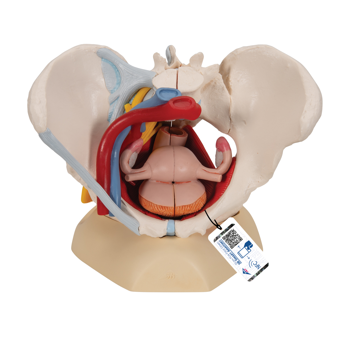
Anatomical Teaching Models Plastic Human Pelvic Models Female Pelvis With Ligaments Vessels Nerves Pelvic Floor Muscles And Organs from www.3bscientific.com (2017, elsevier) should be consulted. Anatomically correct, the language of the text is clear and concise. Visualise your pelvic floor and see exactly what it is, where it's located and why it is important to train this hidden group of muscles. The female pelvis was designed to make the subject of the pelvis understandable to all women. It's located between the abdomen and the legs. This mri female pelvis sagittal cross sectional anatomy title tool is absolutely free to use. Female pelvic applied anatomy by dr shashwat jani dr shashwat jani. It is composed of inlet, cavity, and outlet.
The pelvis's frame is made up of the bones of the pelvis, which connect the axial skeleton to the femurs, and therefore acts in weight bearing of the upper body.
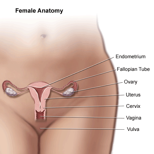
The bony pelvis in normal standing posture transmits the body weight of head, trunk and the upper extremities to the lower extremities. The first step is assessing the mass' site of origin and its location in relation to the peritoneal cavity and the extraperitoneal spaces. Anatomy of the female pelvis. In women, the pelvis houses the uterus, tubes, ovaries and vagina. • pelvis begins at the iliac crests and ends at the symphysis pubis. It is composed of inlet, cavity, and outlet. The female bony pelvis is divided into: Differentiate the different types of the female pelvis. I'm so glad you're joining me as i take you on a user friendly and useful guide to understanding the female pelvis, her relationships and how to assess her! In female it is adapted for child bearing. This mri female pelvis sagittal cross sectional anatomy title tool is absolutely free to use. This mri female pelvis axial cross sectional anatomy title tool is absolutely free to use. (2017, elsevier) should be consulted.
Terms in this set (72) abdominal aorta. Pelvic diaphragm —the term pelvic diaphragm refers to the levator ani muscle and its covering fasciae, both the superior fascia and the inferior fascia. Describe the boundaries and subdivisions of the pelvis. The female bony pelvis is divided into: Visualise your pelvic floor and see exactly what it is, where it's located and why it is important to train this hidden group of muscles.
Female Pelvic Anatomy Artwork Stock Image C010 7098 Science Photo Library from media.sciencephoto.com The female pelvis was designed to make the subject of the pelvis understandable to all women. The obstetrical anatomy of a typical female pelvis is best considered as one unit. Anatomy of the female pelvis. Gynecoid/ genuine pelvis, the brim is round, more wider, and both ischial spines are less prominent this allows easy baby delivery. This is so a baby can pass through the pubic outlet, the circular hole in the middle of the pelvic bones, during childbirth. The pelvis is the lower part of the torso. This mri female pelvis sagittal cross sectional anatomy title tool is absolutely free to use. Female pelvic applied anatomy by dr shashwat jani dr shashwat jani.
Satheesha nayak b, professor at department of anatomy of melaka manipal medical college mmmc (mahe), manipal, india.
Gynecoid/ genuine pelvis, the brim is round, more wider, and both ischial spines are less prominent this allows easy baby delivery. 1 chapter 1 applied clinical obstetric anatomy nasrat1949. Pelvic diaphragm —the term pelvic diaphragm refers to the levator ani muscle and its covering fasciae, both the superior fascia and the inferior fascia. The pelvis is the lower part of the torso. Differentiate the different types of the female pelvis. The female pelvis was designed to make the subject of the pelvis understandable to all women. Use the mouse scroll wheel to move the images up and down alternatively use the tiny arrows (>>) on both side of the image to move the images.>>) on both side of the image to move the images. This mri female pelvis sagittal cross sectional anatomy title tool is absolutely free to use. Hi and welcome to my functional female pelvic anatomy course! This mri female pelvis axial cross sectional anatomy title tool is absolutely free to use. This area provides support for the intestines and also contains the bladder and reproductive organs. Describe the components & function of the pelvic diaphragm. The female pelvic bones are typically larger and broader than a male's.
Visualise your pelvic floor and see exactly what it is, where it's located and why it is important to train this hidden group of muscles. Laparoscopic anatomy of the female pelvic region. Over 250 drawings illustrate every important aspect of pelvic anatomy, and show the reader how to perform simple exercises to keep the pelvis and its related structures fit. In women, the pelvis houses the uterus, tubes, ovaries and vagina. Satheesha nayak b, professor at department of anatomy of melaka manipal medical college mmmc (mahe), manipal, india.
Anatomy Of Female Pelvic Area from api.kramesstaywell.com List the arterial & nerve supply list the lymph & venous drainage of the pelvis. In women, the pelvis houses the uterus, tubes, ovaries and vagina. Above the pelvic brim and has no obstetric importance. Terms in this set (72) abdominal aorta. In female it is adapted for child bearing. Get it as soon as wed, may 26. Nerves of the female pelvis medical illustration nerves of the female. It is composed of inlet, cavity, and outlet.
The pelvis's frame is made up of the bones of the pelvis, which connect the axial skeleton to the femurs, and therefore acts in weight bearing of the upper body.
Hi and welcome to my functional female pelvic anatomy course! The female bony pelvis is divided into: Surgical anatomy of the female pelvis by laparoscopy. Describe the components & function of the pelvic diaphragm. The female pelvic bones are typically larger and broader than a male's. Anatomically correct, the language of the text is clear and concise. The obstetrical anatomy of a typical female pelvis is best considered as one unit. The external female anatomy includes the pubis and the vulva. The female pelvis was designed to make the subject of the pelvis understandable to all women. The pelvis is the lower portion of the trunk, located between the abdomen and the lower limbs. It's located between the abdomen and the legs. This area provides support for the intestines and also contains the bladder and reproductive organs. Terms in this set (72) abdominal aorta.
Gynecoid, anthropoid, android, and platypelloid pelvic anatomy. The external female anatomy includes the pubis and the vulva.

0 Comments:
Posting Komentar